A neighbor of mine had a Norway Maple that looked sick, and asked me to take a look at it.
In spite of the grass being lush and green, showing there was sufficient water
and nutrients available,
the leaves were drooping. This was at the end of May, when growth should have
been vigorous. Pictures of some leaves taken from this tree are shown below.
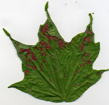
1
In addition to the wrinkled and limp appearance of this leaf, there are also red
blotches present.
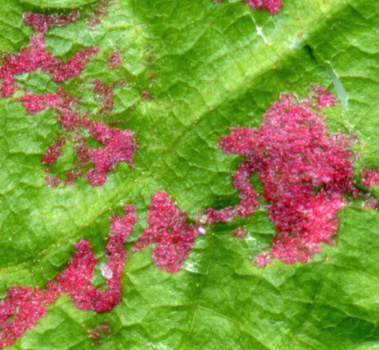
2
A 2400 dpi close-up of one of these red areas shows that it is due to
eriophyid mites, which don't cause serious tree injury.
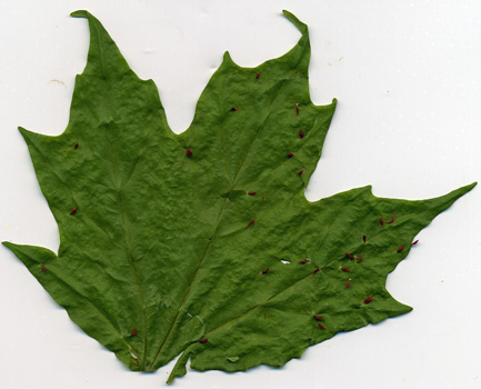
3
Another leaf was also wrinkled. But it had finger-like protrusions
instead.
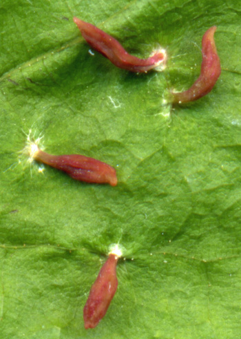
4
This is a 2400dpi close-up of those protrusions (nipple gall?)
5
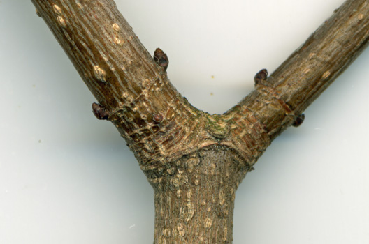
Picture 5 shows a close-up of a branch junction. Click on the picture to zoom
in, and you will see numerous tan particles right at the junction area. Inset
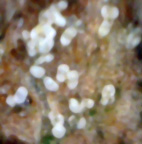
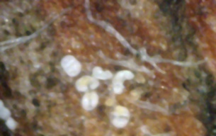
pictures 6 and 7 were obtained using a 400x digital microscope and shows that
these particles are actually spores of white canker that are sapping the tree of
energy. The weakened tree probably makes the tree susceptible to other attacks,
such as mite infestations.
6
7
Having gained more experience in analysis, I returned to this tree in mid-October 2008 to again
gather some branch and leaf samples. However, during the summer this tree had been sprayed
with a fungicide to ward off disease. The following pictures were obtained using a 400x
digital microscope.
8
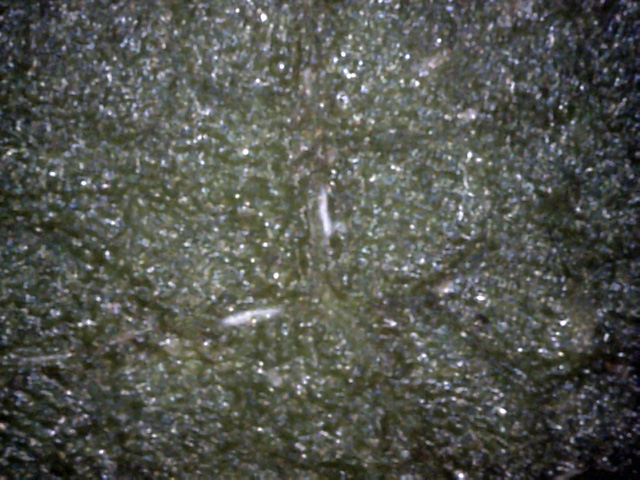
This leaf top is unusually clean - there are just two small traces of canker here
(two white streaks).
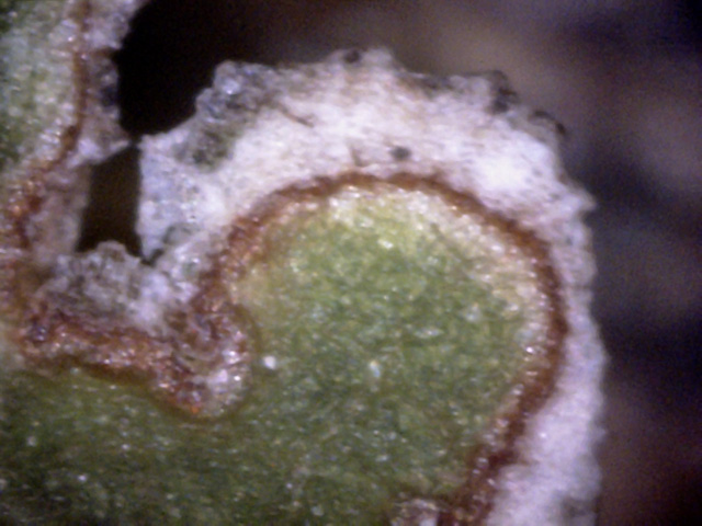
9
This top view is the edge of a leaf tear, due to mechanical damage or disease.
It looks like canker has taken over the dead part.
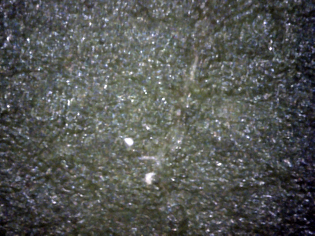
10
While the leaf top is unusually clean, there is a hint that some canker lies just below the surface.
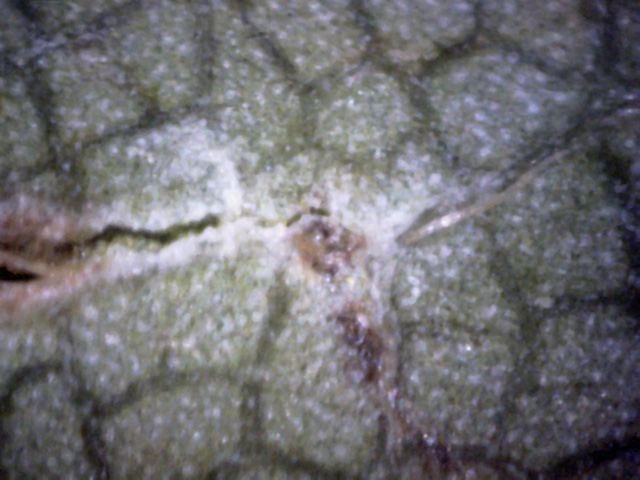
11
The leaf bottom is also unusually clean.
Here is the edge of a tear. There is also a hypha to the right of it.
The white material looks like, but may not be, white canker.
The following five pictures are all leaf cross-section views, obtained using a
400 power digital microscope.

12
There is a pillar of white canker running from to the of the leaf to the bottom.
Two pieces of canker are also hanging off the bottom of the leaf.
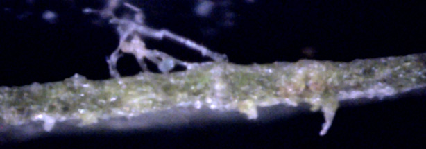
13
A tangled hypha on the top of this leaf indicates the probable presence of white canker.
A few pieces also hang off the bottom of the leaf.

14
This picture shows that much of the white canker that is present lies within the leaf
rather than at or on the surface of it.
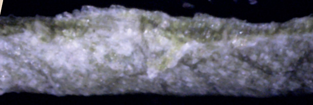
15
There is a high density of white canker just to the left of center, both on the leaf surfaces
and within the leaf.
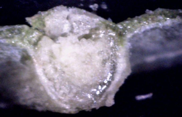
16
This is a leaf vein. The pith is in the center.
But it appears as if the top of this vein has been infected with white canker.
The next three pictures are twig cross-section views, obtained using a
400x digital microscope.
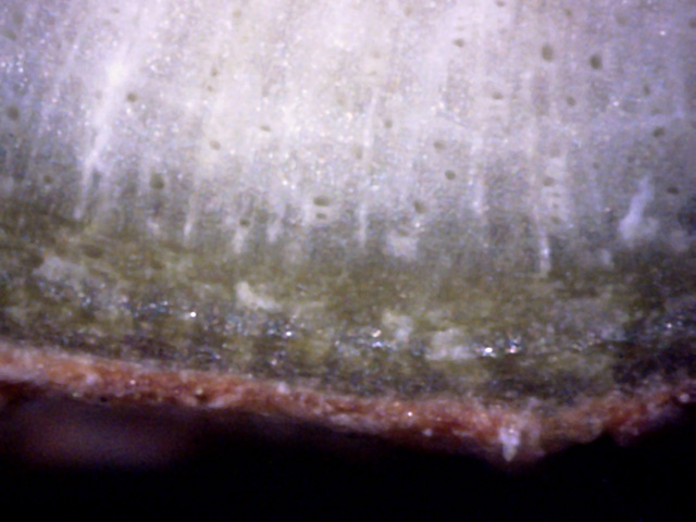
17
It appears the fungicide spraying helped, as this area of the bark is relatively
clean and healthy - the bark and phloem are intact, with only traces of white canker.
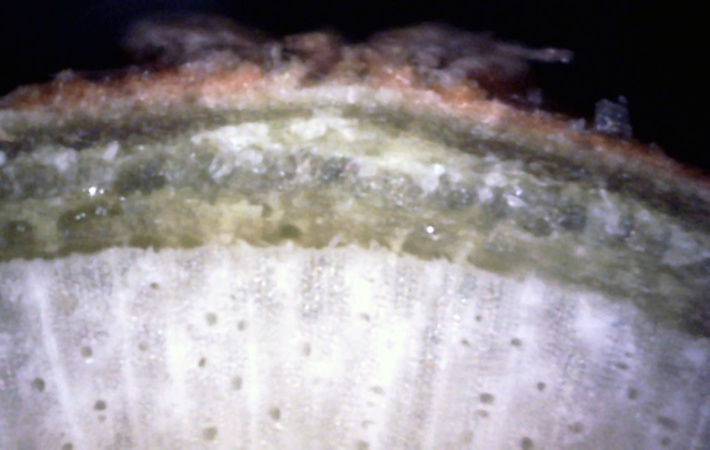
18
The phloem is distorted here, with white canker growing in its outer part and within the bark.
It looks like some hyphae are also pushing through the bark.
Note the slight bulge in the bark developing at this point.
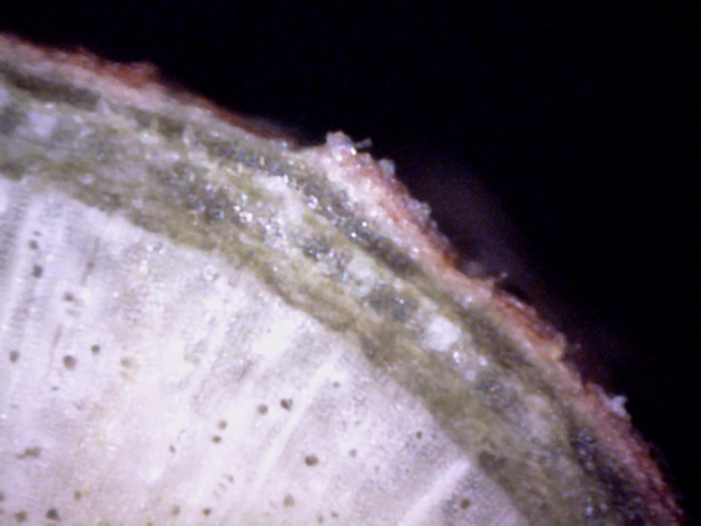
19
Another area where white canker is developing in the outer phloem and the inner bark,
pushing a piece of outer bark out.
In summary, while white canker is present in this tree, the summer fungicide
spraying has reduced the infection significantly - to the point where only
microscopic views provide useful diagnostic evidence. The bottoms and tops of the
leaves give only a hint of infection. The leaf cross-sections more clearly show
signs of infection. But once again, the clearest sign of white canker infection
is seen in twig cross-sections, where the white canker is concentrated at the
outer phloem and the inside of the bark. This is the area high in nutrients.
My neighbor's 25' tall Norway maple became diseased around 2003, and was the first tree in this area to show
symptoms of White Canker. In retrospect, I suspect the infection came in via a
load of infected bark mulch. At this point in time I had no microscope to
confirm the diagnosis using leaf and twig cross-sections, as well as the
presence of White Canker spores. That came several years later.
 - diseased - 800dpi-A.jpg)
1
The dark brown spots on this leaf seem to indicate that it has a bad case of anthracnose.
However, the puckered and dry appearance of the leaf are symptoms associated with White Canker.
The two diseases together are severely weakening the tree, causing significant leaf-drop.
2
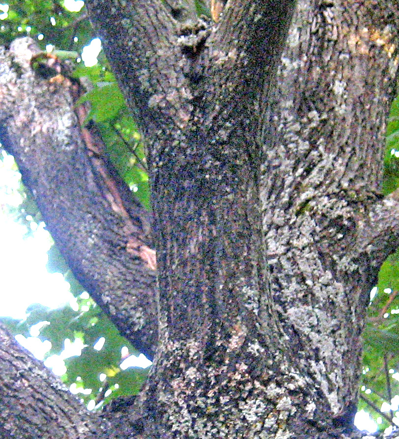
From a distance, the most obvious sign of a White Canker infection is bark fissures.
There may be many small uniform fissures, or one large fissure, or a combination of the two,
depending upon the type of tree. Here a branch at the 9 foot level has developed a
large fissure in the bark. Such a branch usually dies within a year or two.
3
→
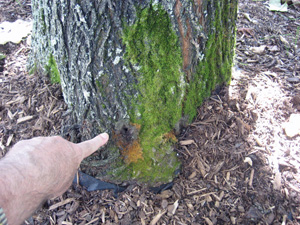
Maple and oak trees infected with White Canker sometimes produce a small hole through which
a watery light-brown liquid drains out. The liquid is odorless. Here my finger is pointing to such a hole.
Notice that the moss below it is discolored. In addition, notice the bark fissures in the upper'right
of this picture (blue arrow).
In summary, while the diagnostic evidence for White Canker is somewhat limited,
it is still strong enough to build a solid case for White Canker. Specifically,
while the puckered leaf provides a strong hint, the weeping bark along with several
characteristic bark fissures provides evidence strong enough to implicate White Canker
as the cause of this tree's decline.
This is the same tree several years later. During this time it was treated with
a fungicide. While each treatment was successful, it only lasted about a month. At
this point in time (early June 2008), no fungicide treatment had been applied
this season, and the tree was again in decline. Now, however, my list of
diagnostic signs had grown to include spore density locations, spore appearance,
and specific White Canker growth locations.
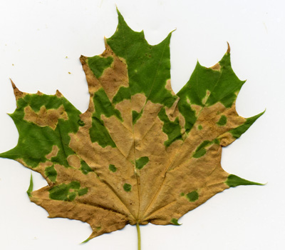
1
The lack of a fungicide treatment during the first part of this summer has caused many of the
leaves to turn color and fall off. This picture shows one of these leaves.
A microscopic look at the leaf showed some white and yellow spores, but not many of them.
It's possible that this tree is suffering from white canker and anthracnose.
2
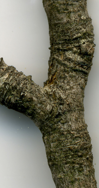
This picture shows a small diseased branch that was near death.
It's easy to see why this branch is sick - the bark is loaded with pale yellow
white canker spores.
3
→
←
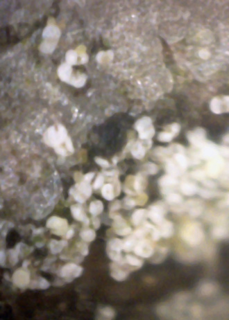
This is a microscopic close-up of one of these spore patches.
Several white spores at the top of the picture still have their embryonic white tissue
between their lobes, indicating that the spores are under construction (blue arrows).
4
→
↓
→
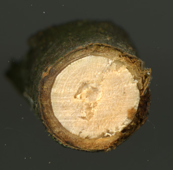
Since the branch shown in picture 2 was only about 3/4 " thick, I used a side cutter
to clip off a clean end, and then scanned it at 2400 dpi, as shown here.
At first glance, there is nothing unusual here.
But look closely (click on the picture) at the upper-right junction of the xylem and phloem (blue arrow).
The tissue layer there has thickened and turned into a waxy-looking substance.
The same thing has happened to the bottom (blue arrow).
Furthermore, the phloem under the bark in these two areas has developed voids, with a void also
forming between the phloem and the outer bark. Continuing on, the bark between these two areas is so
badly diseased that it has decayed and turned a dark brown. In the middle of this decayed area the
bark has split, turning the xylem underneath it brown in color (red arrow).
This cross-section is an excellent tutorial on the damage done by white canker.
5
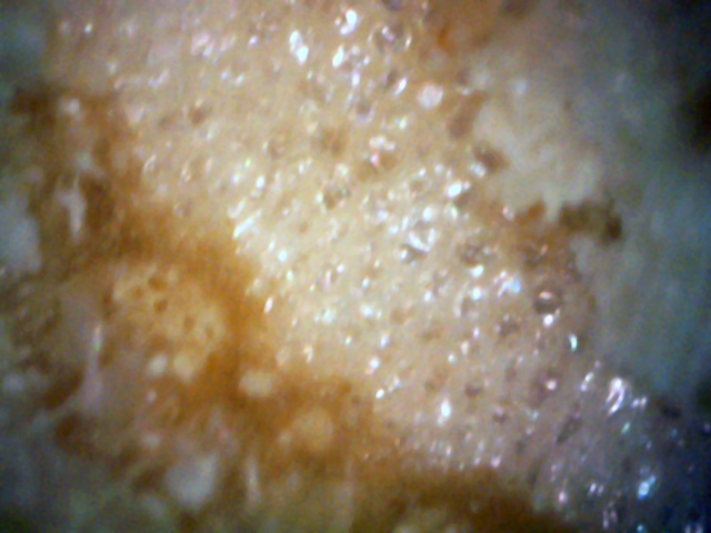
This is a microscopic view of the pith at the center of the branch. It has a frothy, bubbly look.
This is normal. So far, I've never seen white canker infect the pith.
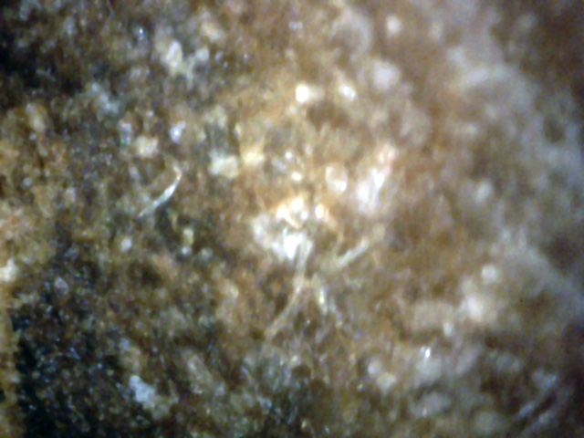
6
When I examined some "good" surface bark with a microscope, I saw what appeared to be
white spores just under the surface, along with a few hypha, as shown above.
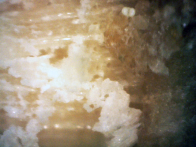
7
Picture 4 seems to show that the outer tree rings have been replaced by a waxy substance.
This picture shows what this substance looks like when viewed with a 400x microscope.
It appears as if white canker material is replacing the original woody tissue.
The wrinkled leaf gave a hint of white canker infection, while its discoloration hinted at
anthracnose. The abundance of light yellow spores on the bark was a strong piece of evidence, confirmed
by the microscope picture of the spores themselves, which were identical in shape and size to
white canker spores. Finally, a 2400 dpi scan of a branch cross-section showed how this branch died:
the white canker consumed the phloem layer, preventing nutrient transport. The voids created under the
bark cut off nutrients to the bark, causing it to decay and split.
Several months later, the leaf drop of this tree accelerated, and I decided to
take another look at it.
Picture 1 shows a typical leaf. The leaves have a dry, wrinkled look, as if they lack water.
Yet, the past several months have been exceptionally wet due to a persistent rainy weather system.
(At the end of the year, the weatherman said this was the wettest year on
record! So, lack of rain is definitely not a problem here.)
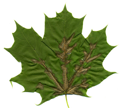
1
This leaf appears to have anthracnose, but other evidence
hints that anthracnose is likely a secondary disease.
Suspecting that there may be an underlying cause to this tree's problems, I used
a 400x digital microscope to examine other parts of the tree.
Three leaf and twig pictures are shown below.
The blue lines are 50 micrometer scale bars.
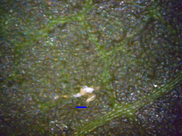
2
The leaf top surface is generally clean, with only a hint of white canker.
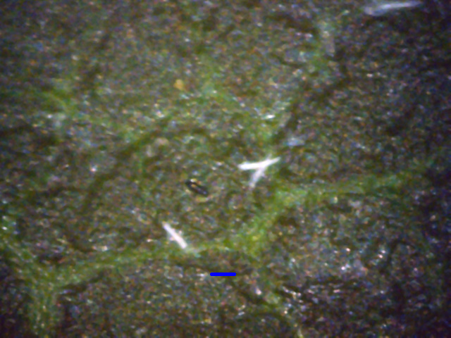
3
Likewise, the leaf bottom only shows hints of a white canker infection.
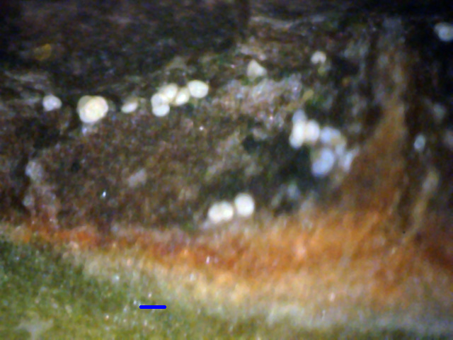
4
This twig shaving view shows hints of trouble: there are numerous spores on the
bark's surface, and underneath the bark there is a white substance above the
green cortex.
I used the same 400x microscope to examine some 3/16" twig cross-cut views next,
with the results shown below.
5
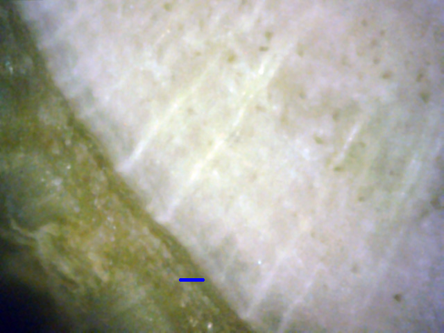
This picture shows a healthy area - the white xylem joins the green phloem with
no air gaps to be seen. Look closely, however, and there are signs of white
canker just beginning to infect the phloem.
6
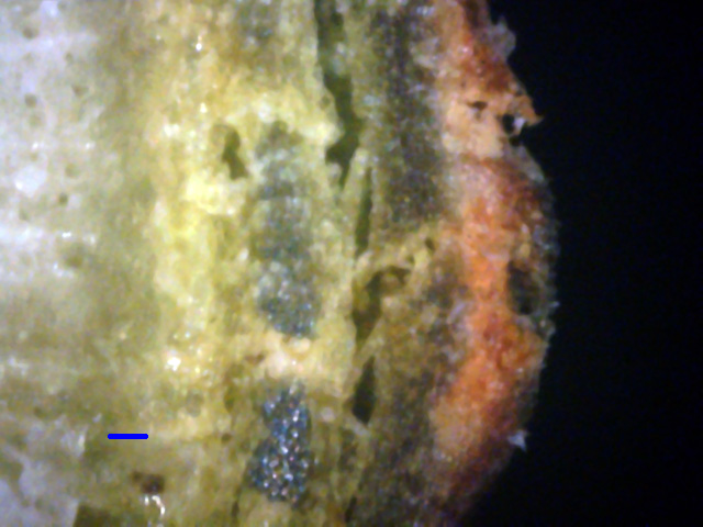
Far more common is the disorganized growth shown here, where the rich green of
the phloem is being replaced with white canker material that ranges from gray to
yellow. In fact, the yellow-gray canker has started to invade the snow-white
sapwood.
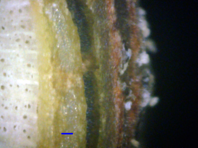
7
This is an excellent view of how the canker formed layers of waxy
material. The bark on the right of this diseased area is growing spores.
The next pictures show several other areas under the bark.
The green phloem layer has pretty much been destroyed, being replaced with a variety of
cankerous material. Compare these pictures to the relatively healthy tissue
shown in picture 5.
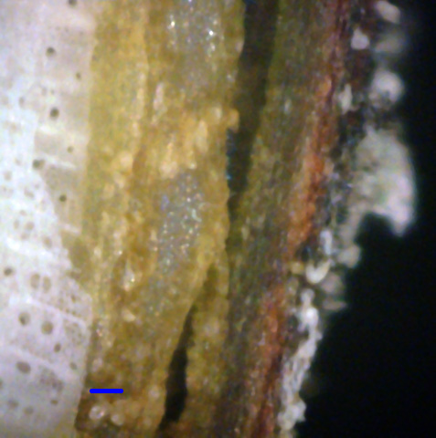
8
This picture shows that the canker has almost completely destroyed the phloem,
replacing it with a waxy material and air gaps. Furthermore, the canker is now
growing into the xylem and is growing particles on the surface of the bark.
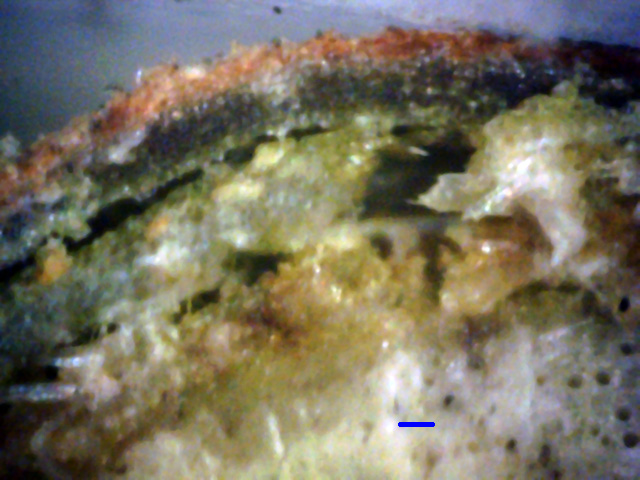
9
An area of mass destruction caused by white canker. The phloem layer is
virtually unrecognizable, having been almost totally consumed by white canker,
which here appears as light gray in color.
In Summary, while the leaves on this tree may indicate an anthracnose infection, the tree in fact
has a much more severe white canker infection at its root. The evidence of this is only barely hinted
at by the dryness and curling of the leaves. Even a microscopic leaf surface examination shows little
evidence. A leaf cross-section gives stronger evidence of white canker. But by far the best evidence
of white canker is a 400x microscopic twig cross-section, where the ongoing destruction of the
tree's phloem tissue is plainly evident. Since the phloem layer carries the nourishment for the
tree, the white canker is effectively choking the tree of the food needed to live and fight off
disease.


 - diseased - 800dpi-A.jpg)


